Scanning Electron Microscopy
Browse 2,068 authentic scanning electron microscope stock photos, high-res images, and pictures, or explore additional scanning electron micrograph or electron microscope micrographs stock images to find the right photo at the right size and resolution for your project. scanning electron micrograph electron microscope micrographs microscope slide

Biology 130 Lab 3 Electron Micrographs
Currently, the machine learning (ML)-based scanning transmission electron microscopy (STEM) analysis is limited in the simulative stage, its application in experimental STEM is needed but challenging. Herein, we built up a universal model to analyze the vacancy defects and single atoms accurately and rapidly in experimental STEM images using a full convolution network. In our model, the.
Electron microscopy
In this paper, we present the first publicly available human-annotated dataset of images obtained by the Scanning Electron Microscopy (SEM). A total of roughly 22,000 SEM images at the nanoscale.
Scanning Electron Microscopy Gallery Center for Microscopy and Imaging
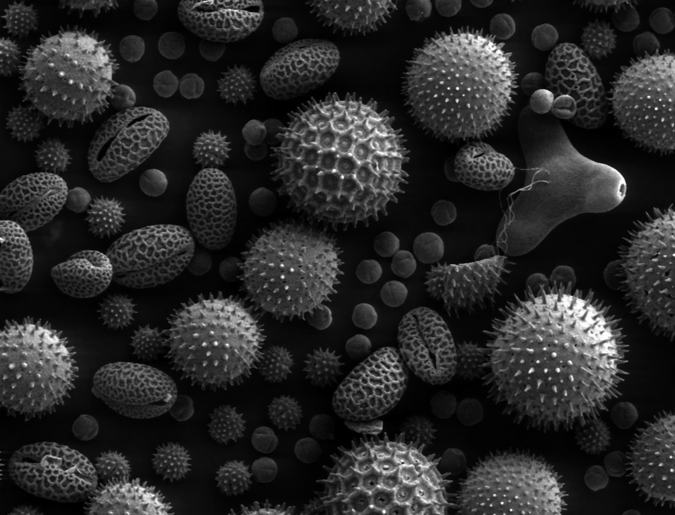
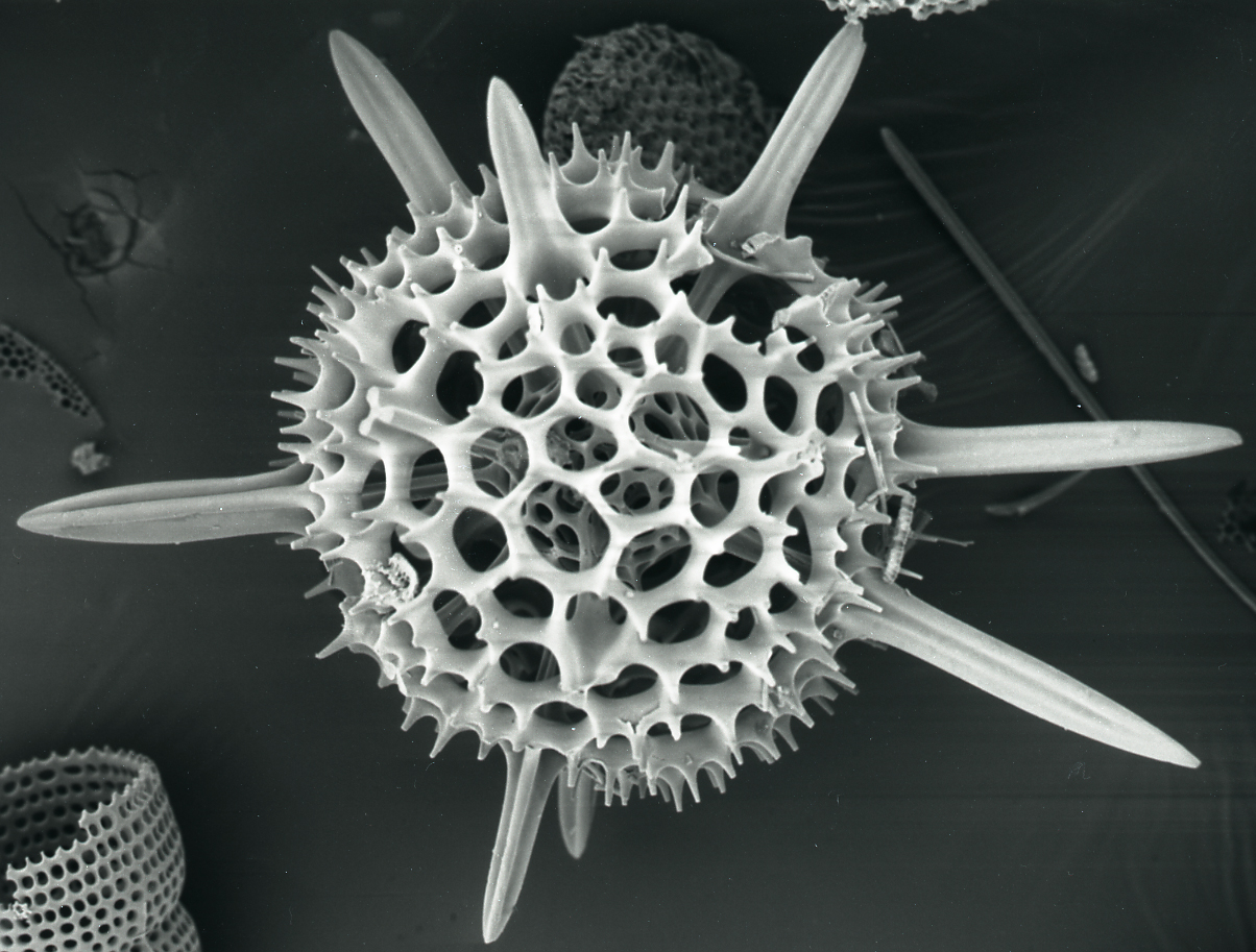
A scanning electrode microscope ( SEM) is a type of electron microscope that produces images of a sample by scanning the surface with a focused beam of electrons. The electrons interact with atoms in the sample, producing various signals that contain information about the surface topography and composition of the sample.

Scanning electron microscope (SEM) Definition, Images, Uses
Browse 5,067 authentic electron micrograph stock photos, high-res images, and pictures, or explore additional electron micrograph nucleus or electron micrograph mitochondria stock images to find the right photo at the right size and resolution for your project. electron micrograph nucleus electron micrograph mitochondria
Scanning Electron Microscopy Images Central Microscopy Research Facility
However, on March 20, 2021, Muller again led a team that has beaten its own record with what is described as an electron microscope pixel array detector (EMPAD) that has an even more.
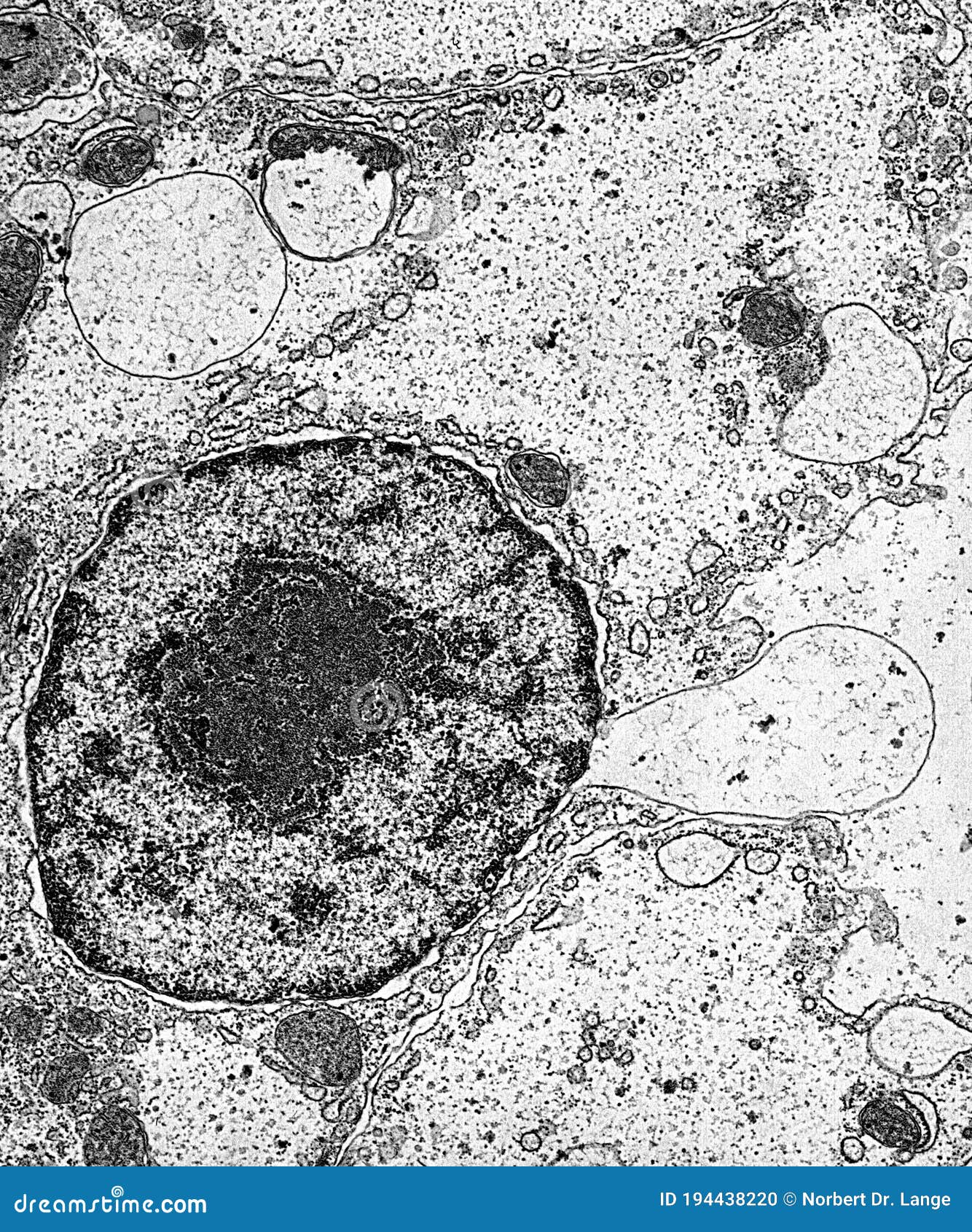
Cell Nucleus and Organelles Under the Electron Microscope Stock Photo
By Anna Blaustein Image shows an electron ptychographic reconstruction of a praseodymium orthoscandate (PrScO 3) crystal, zoomed in 100 million times. Credit: Cornell University September 2021.
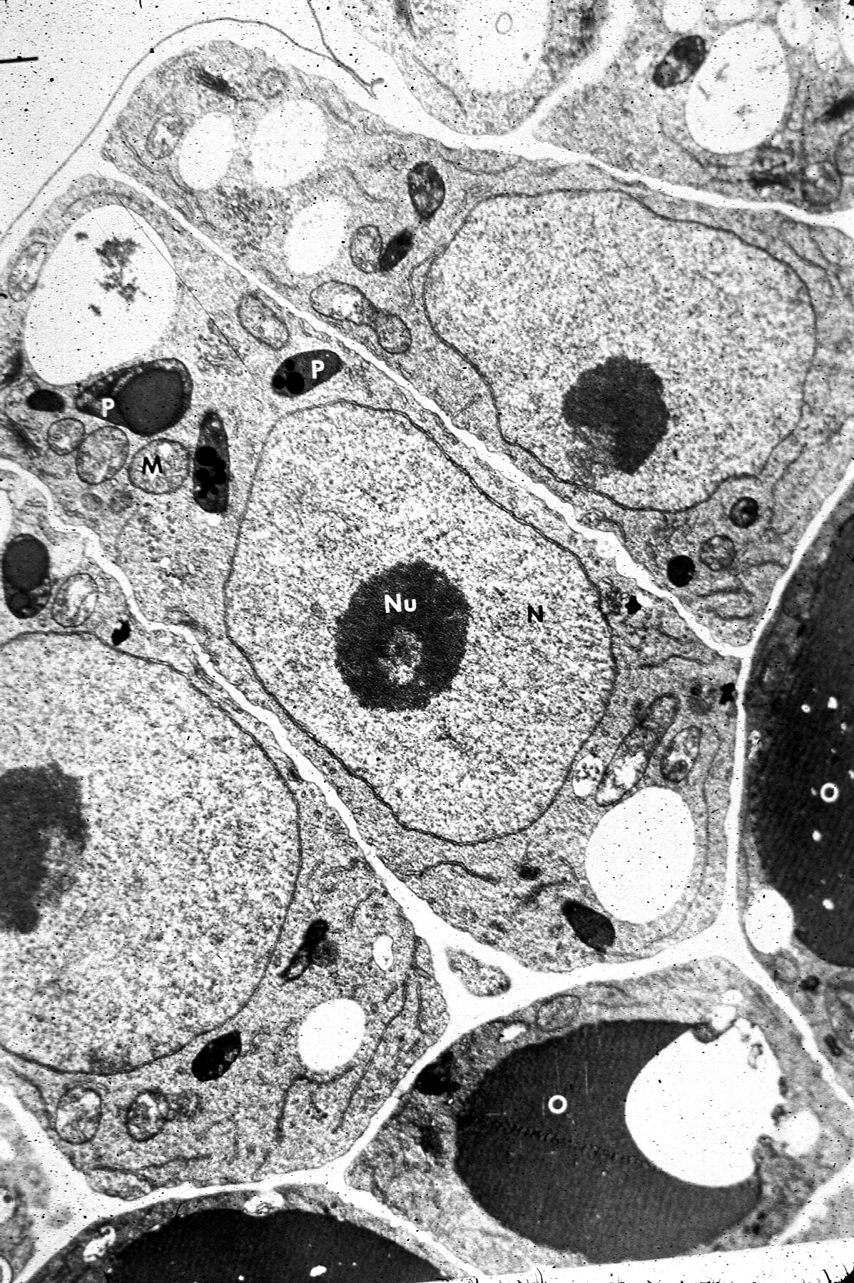
Electron Micrograph
Electron Microscope Photos and Premium High Res Pictures - Getty Images AI Generator Images Browse millions of royalty-free images and photos, available in a variety of formats and styles, including exclusive visuals you won't find anywhere else.
New Electron Microscopy Technique Offers First RealTime Look at
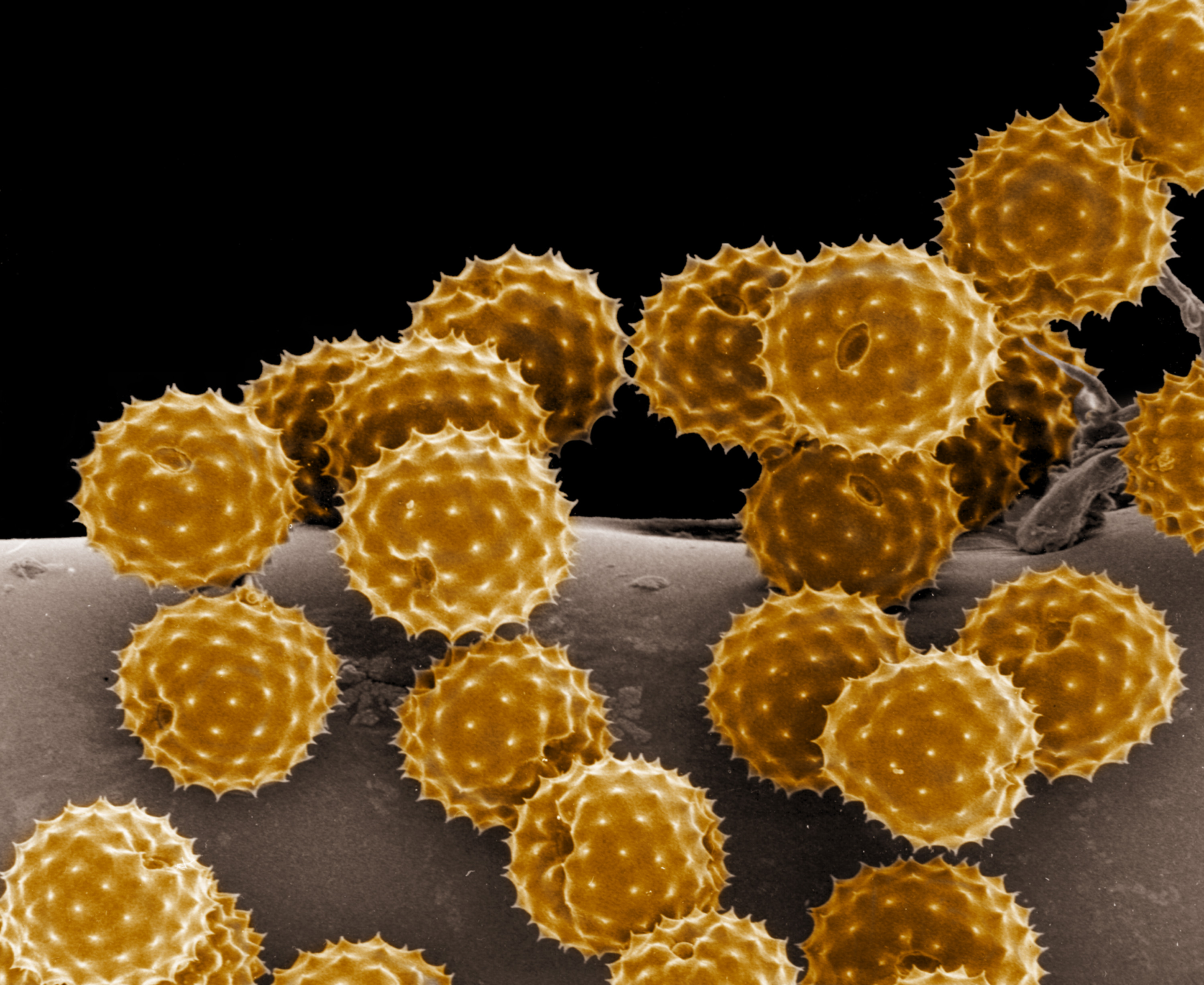
The pollen grains contain a male gamete (reproductive cell) that is intended to fertilise an egg or ovule (female gamete). This will initiate the formation of a seed for a new plant. Coloured scanning electron micrograph (SEM), magnified x250 when printed at 10cm wide. electron microscope stock pictures, royalty-free photos & images
Biology 130 Lab 3 Electron Micrographs
2,998 Electron Microscope Images Stock Photos, High-Res Pictures, and Images - Getty Images Images Creative Images Browse millions of royalty-free images and photos, available in a variety of formats and styles, including exclusive visuals you won't find anywhere else. See all creative images Trending Image Searches Happy New Year New Year Family
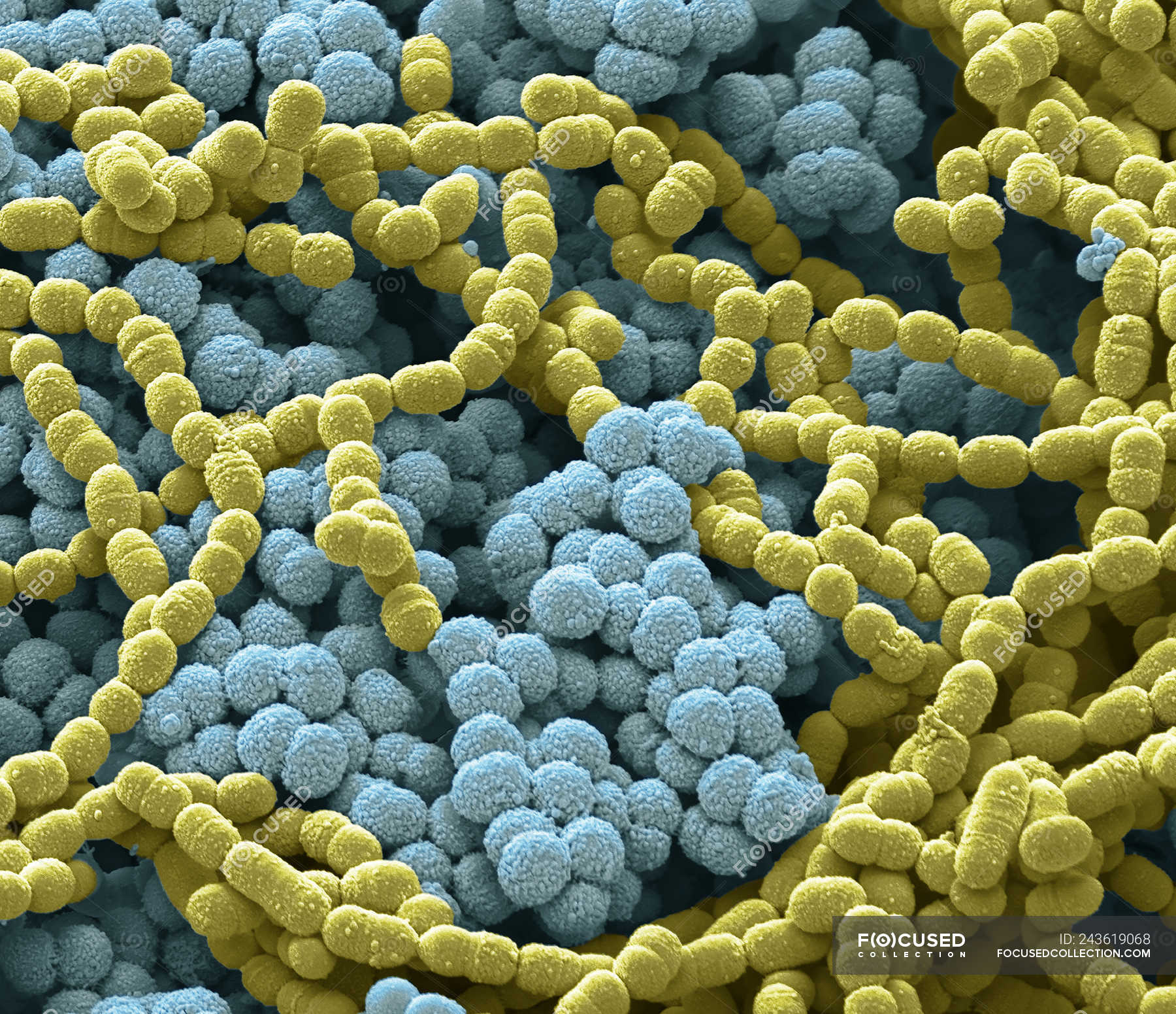
Scanning electron micrograph of bacterial culture from sputum
Electron microscopy (EM) uniquely visualizes cellular structures with nanometre resolution. However, traditional methods, such as thin-section EM or EM tomography, have limitations in that they.

30 Of the Most Amazing Images from Electron Microscopes Electron
Bringing color to electron microscope images is a tricky problem. It could plausibly be said that color doesn't exist at that scale, because the things imaged by an electron microscope are.

Scanning Electron Microscpy Photography by Robert Berdan The Canadian
Find Electron micrograph stock images in HD and millions of other royalty-free stock photos, illustrations and vectors in the Shutterstock collection. Thousands of new, high-quality pictures added every day.
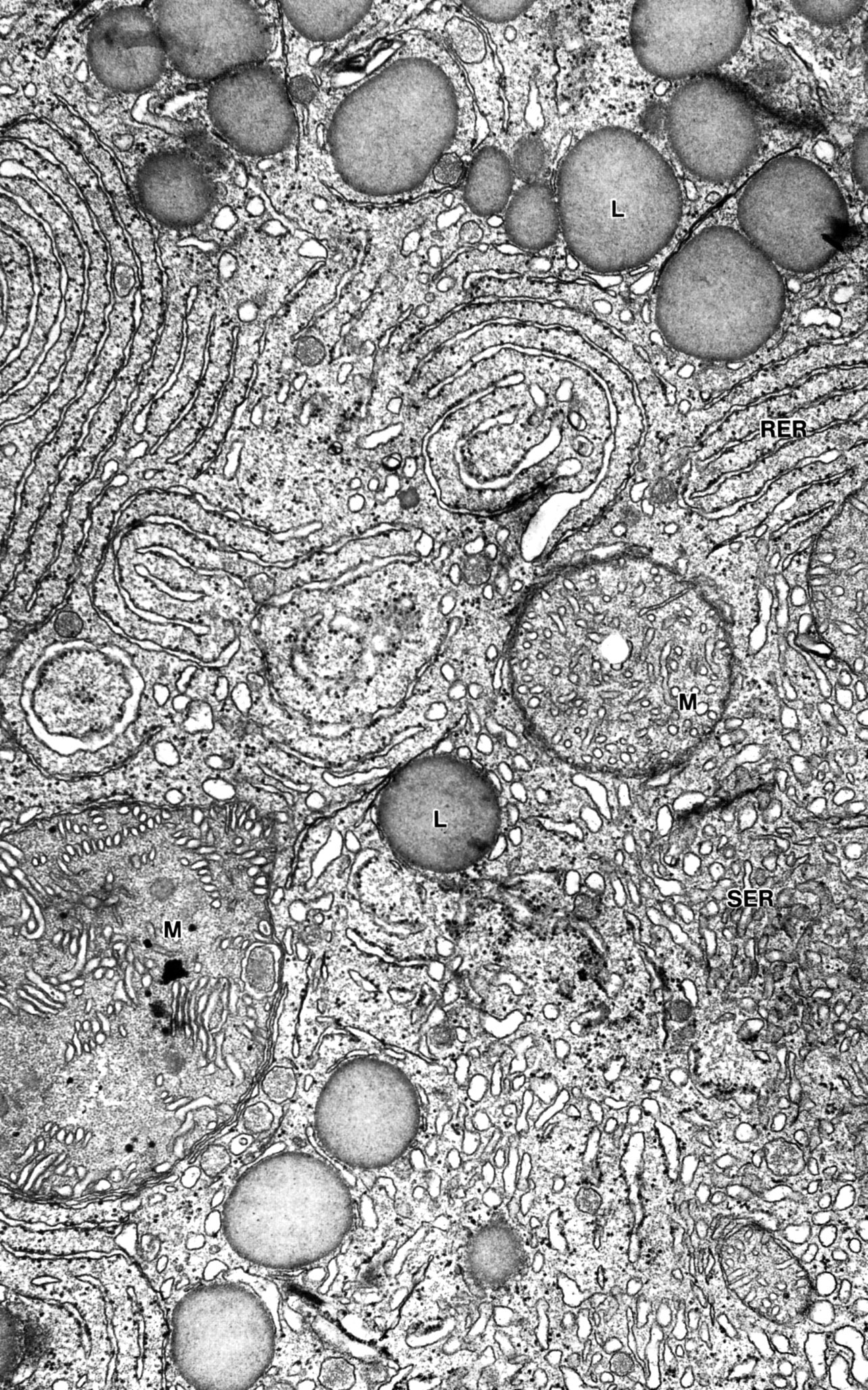
Transmission electron micrograph of an animal cell Stock Image G450
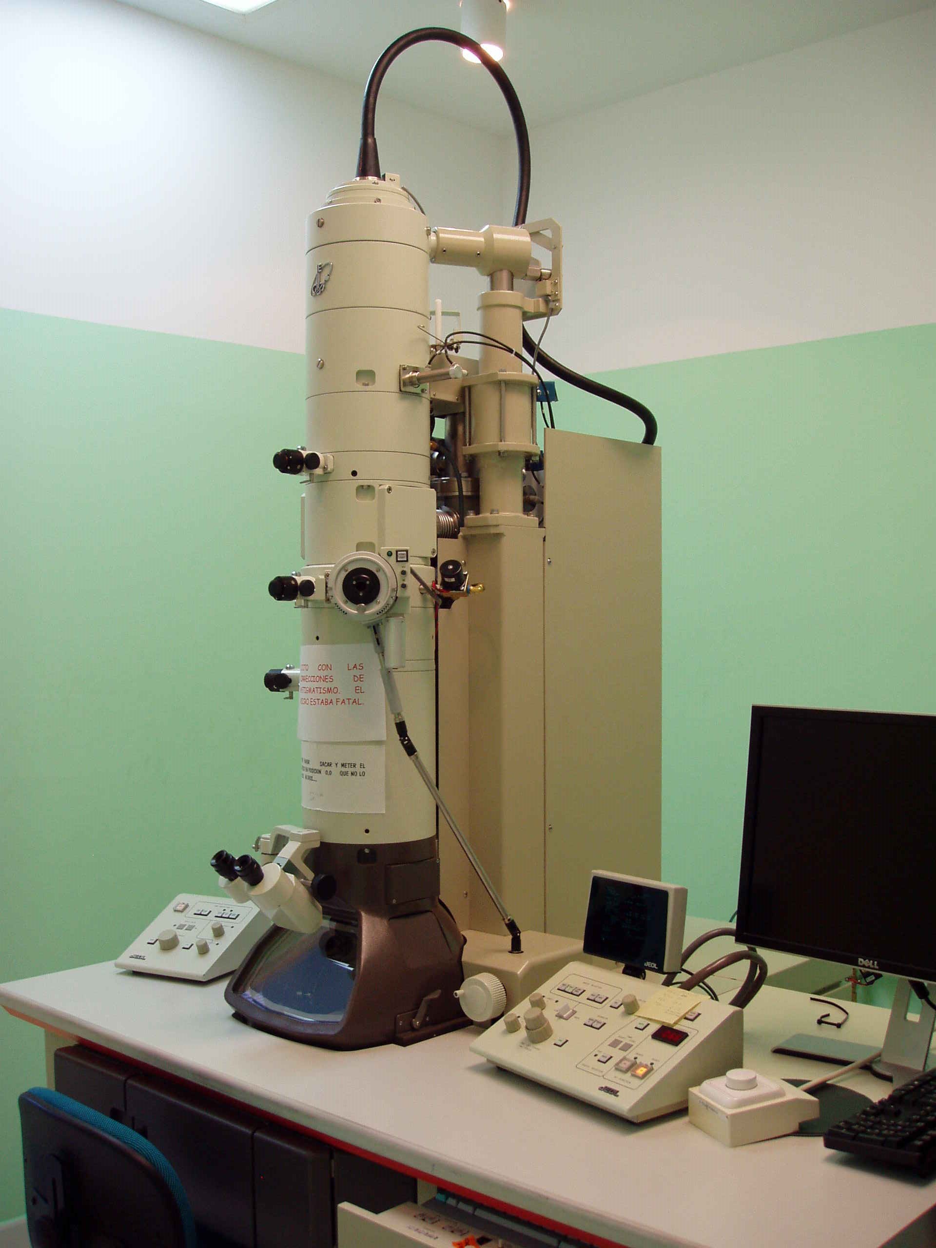
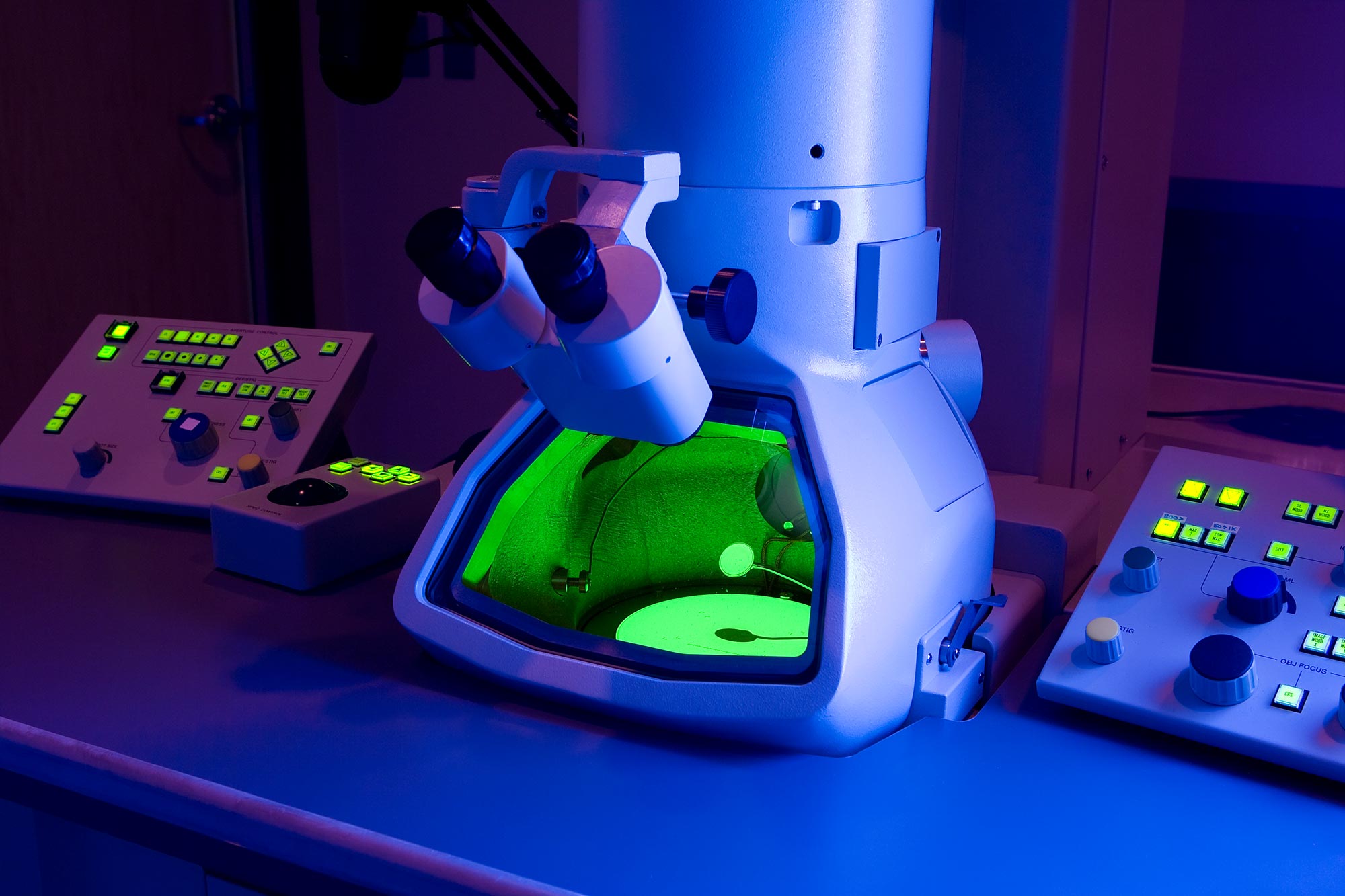
An electron microscope is a microscope that uses a beam of electrons as a source of illumination. They use electron optics that are analogous to the glass lenses of an optical light microscope to control the electron beam, for instance focusing them to produce magnified images or electron diffraction patterns.

How Does A Scanning Electron Microscope Develop Such Breathtaking Images?
Advanced electron microscopy techniques, including scanning electron microscopes (SEM), scanning transmission electron microscopes (STEM), and transmission electron microscopes (TEM), have.
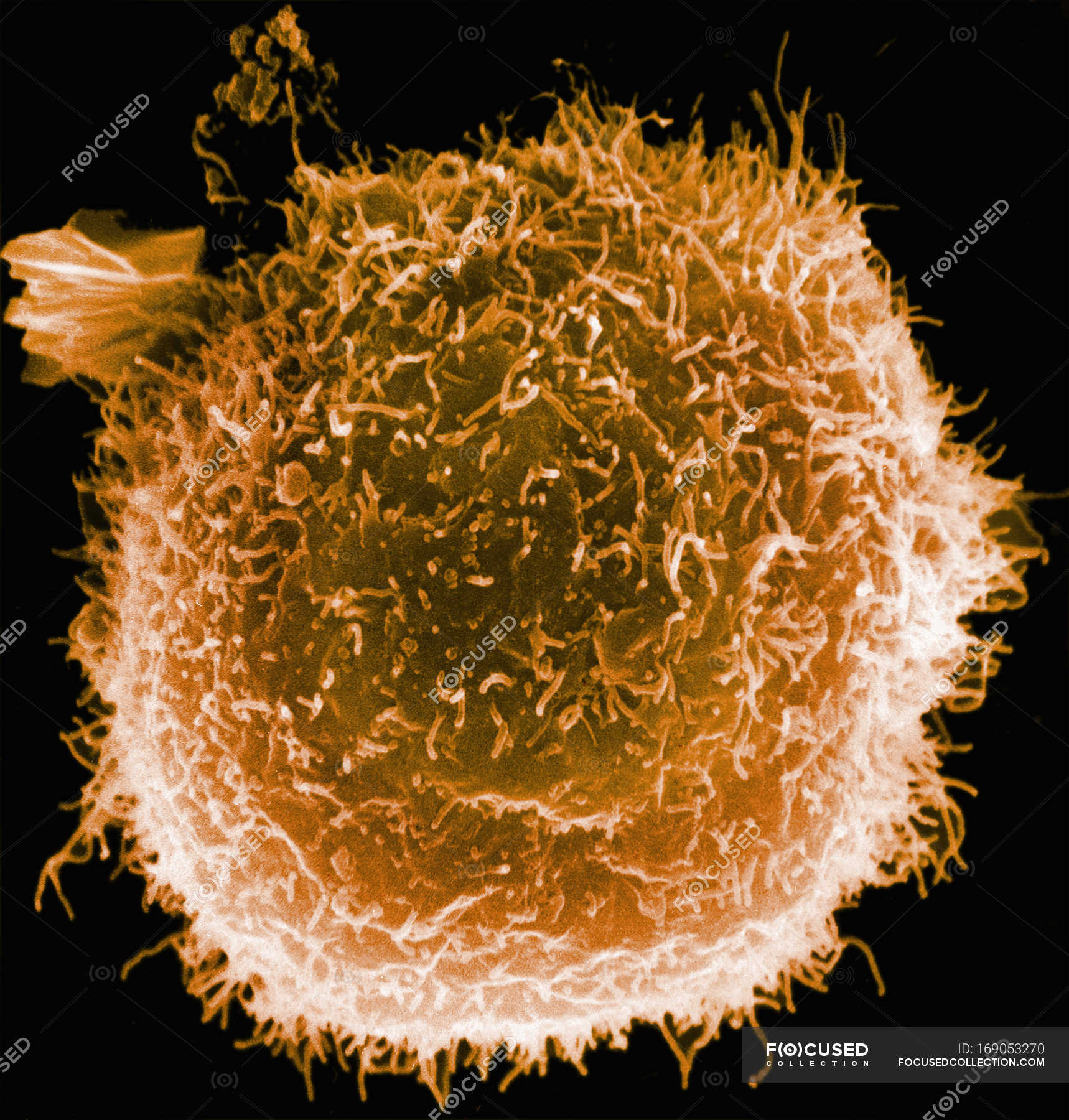
Scanning electron micrograph of human macrophage — biological, White
EMPIAR, the Electron Microscopy Public Image Archive, is a public resource for raw images underpinning 3D cryo-EM maps and tomograms (themselves archived in EMDB).EMPIAR also accommodates 3D datasets obtained with volume EM techniques and soft and hard X-ray tomography.